CHENNAI — The Sudha Gopalakrishnan Brain Centre at the Indian Institute of Technology Madras has launched an unprecedented initiative to construct three-dimensional cellular reference maps of 100 human brains spanning from the foetal stage to an 85-year-old individual. Unveiled recently by Prime Minister Narendra Modi, the project represents a global first in mapping the entire human central nervous system at microscopic resolution across an entire lifespan.
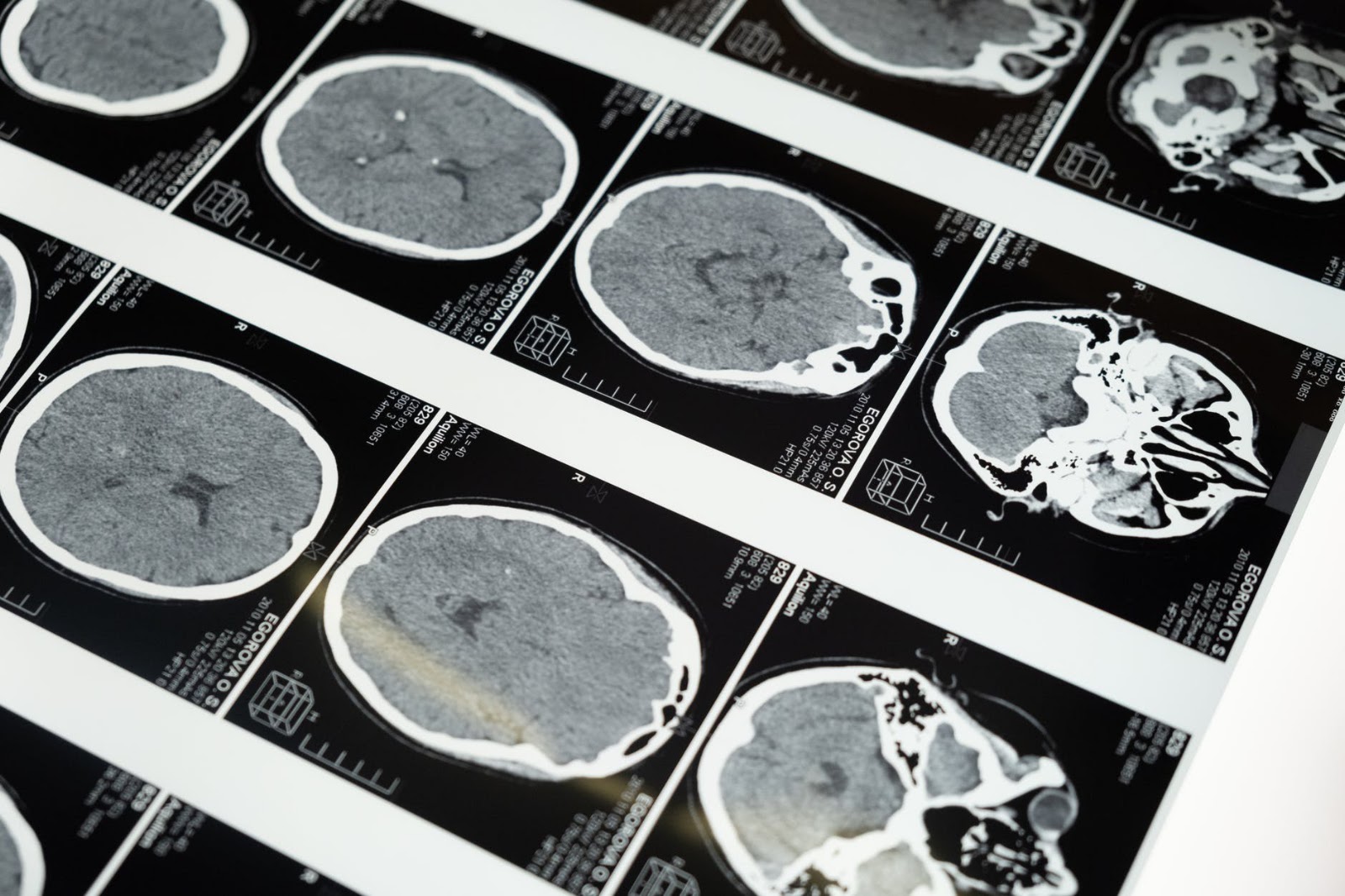
While conventional magnetic resonance imaging (MRI) provides clinical views of living patients at a millimetre scale, it fails to capture the underlying cellular machinery where neurological anomalies originate. By examining the human brain at a scale previously unachieved across human aging, researchers are attempting to bridge a foundational blind spot in clinical medicine. Understanding the organ’s physical transformation across decades could unlock crucial insights into neurodegenerative decline, congenital abnormalities, and ischemic injury.
Industrial-Scale Precision at the Cellular Level
To construct these massive digital models, scientists begin by freezing post-mortem donor brains to preserve their anatomical integrity. Specialist automated instruments, engineered and customized directly by IIT Madras, section the tissue into ultra-thin slices measuring between 10 and 20 microns—roughly one-tenth the width of a single human hair.
The sheer physical scale of this process varies dramatically by developmental phase. Processing a fully grown adult brain demands slicing between 8,000 and 10,000 individual sections. In contrast, reconstructing a developing foetal brain requires roughly 3,000 sections, while a young child’s brain yields approximately 5,000 slices. Every cross-section is digitized under high-powered microscopy before computational frameworks reconstruct the separate images into a coherent, navigable three-dimensional matrix.
"Today, we don’t have a detailed view of how a human brain and its structures look across the lifespan. The brain evolves continuously, unlike other organs. We need a map of how the brain looks at various ages. Only then will we be able to translate this into medicine."
Deciphering Lifespan Transitions and Stroke Pathologies
A primary driver of the project is understanding why specific brain regions become uniquely vulnerable at different chapters of life. Brain architecture in infancy bears fundamentally different structural hallmarks than in middle age or senescence. Establishing a standard cellular baseline allows clinicians to distinguish normal developmental variations from nascent disease phenotypes.
The groundwork for the expanded 100-brain initiative originated with DHARANI, the centre’s foetal brain atlas released in 2025, which has since gained widespread international reference among neurobiologists. Expanding on that foundation, the center recently introduced C-STROKE, an atlas that maps an entire stroke-afflicted human brain. C-STROKE provides an uninhibited, omnidirectional perspective of ischemic damage, giving neuroscientists the ability to inspect damaged tissue alongside healthy surroundings at multiple microscopic depths.
Beyond physical morphology, the research team is conducting extensive RNA sequencing to catalog localized molecular activities. Pinpointing the exact molecular processes operating within specific cellular clusters allows researchers to uncover potential drug targets and understand why identical pathologies manifest with varying severity across different patients.
Petabyte Computations and Global Medical Accessibility
The high-throughput scanning protocols produce petabytes of microscopic imaging data, creating substantial computational hurdles in data processing and user accessibility. To parse this monumental repository, IIT Madras designed a dedicated digital visualisation platform paired with an integrated artificial intelligence research assistant. The AI engine enables scientists to detect complex cellular structural patterns, correlate anomalies, and interrogate dense datasets using natural language queries.
The institutional undertaking bridges historically isolated fields, gathering approximately 200 researchers across life sciences, clinical medicine, computer science, and engineering. The centre is actively collaborating with over ten major healthcare institutions, including Christian Medical College (CMC) Vellore, Kasturba Medical College (KMC), and Saveetha, ensuring clinical relevance directly informs scientific modeling. Several visiting Nobel laureates have highlighted the project’s transformative scientific scope as work that carries Nobel-caliber potential.
IIT Madras has confirmed that the entire repository of completed brain atlases will be placed in the public domain. By delivering an open-access blueprint of human neuroanatomy from womb to late life, the project aims to equip neurosurgeons, pharmacologists, and researchers worldwide with the cellular coordinates required to target some of medicine’s most resilient neurological disorders.
TL;DR
- IIT Madras is mapping 100 human brains from foetus to age 85 at cellular resolution.
- The project slices brains into 10 to 20 micron sections to construct 3D digital atlases.
- Adult brains yield between 8,000 and 10,000 physical slices during the imaging process.
- A dedicated stroke atlas, C-STROKE, was unveiled alongside the broader initiative.
- Over 200 researchers and 10 medical institutions are collaborating on the open-access database.